- Where does AI actually help in spine surgery today?
- What has actually been improved or automated?
- Does it help with imaging studies specifically?
- Can it predict specific outcomes for specific treatments?
- Does it genuinely make the surgeon's work easier?
Had spine surgery and the pain returned? Send your studies for a second opinion.
Send my studiesNot in the way science fiction suggests. There is no autonomous AI performing surgery, and there won't be for the foreseeable future. What actually exists today is narrower and more useful: AI systems that read imaging faster and more consistently, models that estimate a patient's individual risk before surgery, navigation and robotic systems that improve the precision of implant placement, and intraoperative monitoring tools that flag danger to the spinal cord in real time. Each of these is a tool that supports a specific decision — not a replacement for the surgeon making it.
AI in spine surgery today is best understood as four separate categories of tool — imaging/diagnosis, outcome prediction, surgical precision (navigation and robotics), and intraoperative safety monitoring — each at a different level of maturity, rather than a single technology. (Benzakour et al., 2023; Hornung et al., 2022)
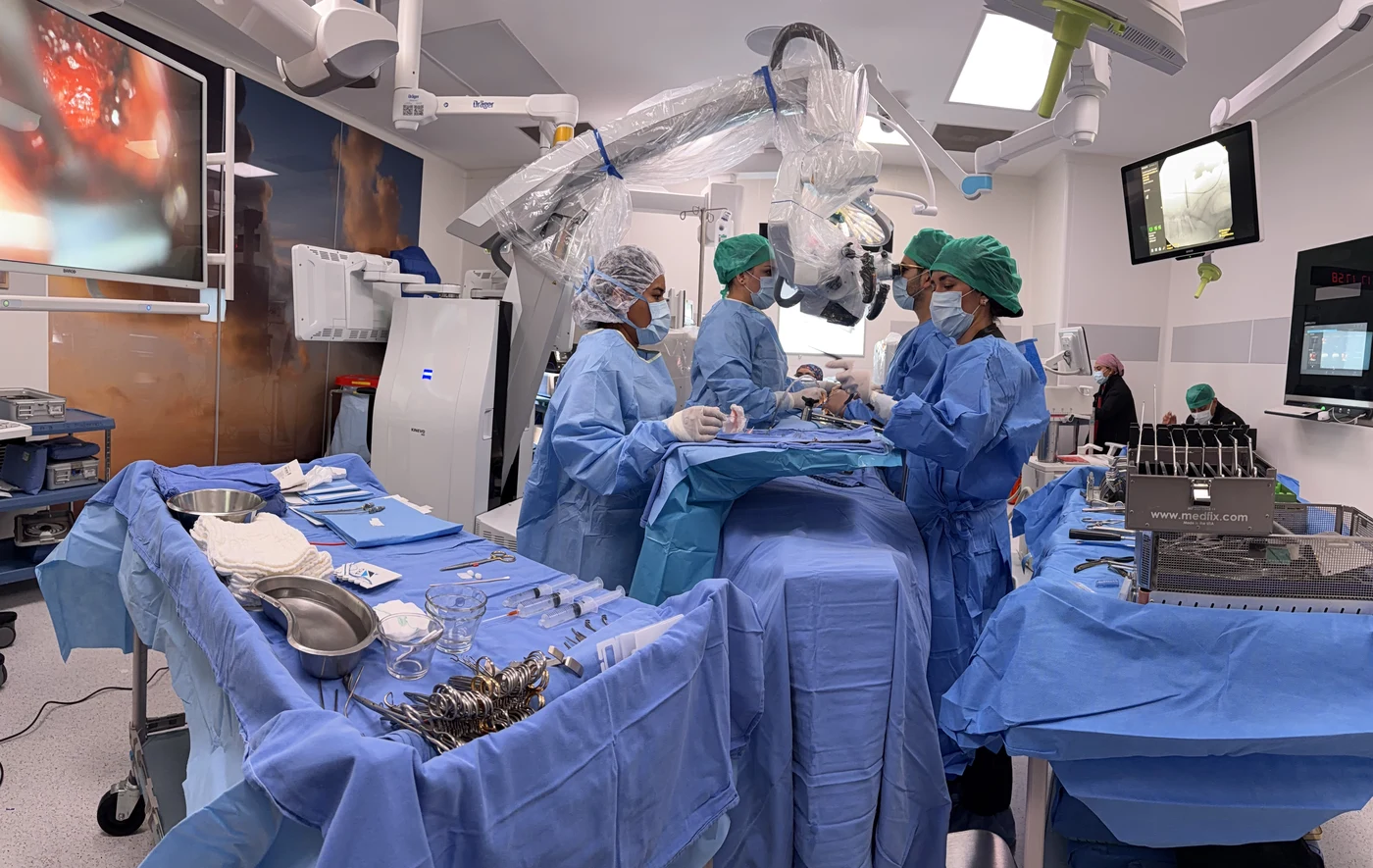
The most mature, clinically deployed application is automated pedicle screw trajectory planning combined with intraoperative navigation and robotic guidance — technology that has moved from research to routine use in many centers. A meta-analysis of nine randomized controlled trials found robot-assisted pedicle screw placement to be more accurate than freehand technique. (Li et al., 2020) Automated image segmentation — outlining vertebrae, discs, and spinal canal boundaries on MRI or CT without manual tracing — has similarly matured to the point of routine research and, increasingly, clinical use. (Jamaludin et al., 2017; Song et al., 2023)
| Application | Maturity level |
|---|---|
| Robot/navigation-assisted pedicle screw placement | Clinically established |
| Automated spinal imaging segmentation | Mature research, growing clinical use |
| Fracture and stenosis detection on imaging | Validated in multiple studies, not yet standard of care |
| Individualized outcome/complication prediction models | Active research, limited clinical deployment |
| Finite element / biomechanical modeling for surgical planning | Emerging, mostly research settings |
| Autonomous surgical decision-making | Does not exist |
This is the area with the deepest evidence base. Deep learning models have been trained and validated to detect lumbar spinal stenosis on CT and MRI (Li et al., 2024; Suzuki et al., 2024), classify osteoporotic vertebral compression fractures on plain radiographs (Shen et al., 2023; Burns et al., 2016), identify cervical spine fractures on CT (Esfahani et al., 2023), and predict curve progression in adolescent idiopathic scoliosis from an initial clinic visit X-ray (Wang et al., 2021). A platform combining natural language processing with imaging has also been developed to automatically classify lumbar disc herniation findings, reducing the manual work of correlating radiology reports with imaging findings. (Wang et al., 2024; Dong et al., 2024)
These tools do not replace radiologist or surgeon interpretation — their demonstrated value is consistency (the same finding read the same way every time) and speed at scale, which matters most in high-volume screening contexts and in flagging cases that need urgent human review.
This is one of the most active areas of research, and results are genuinely promising, though mostly still confined to research settings rather than routine bedside use. Machine learning models have been developed and internally validated to predict adverse events after spine surgery in general (Han et al., 2019), discharge disposition and early unplanned readmission after spinal fusion (Goyal et al., 2019), complications following posterior lumbar fusion using neural networks (Kim et al., 2018), sustained opioid use after both lumbar disc herniation surgery and anterior cervical discectomy and fusion specifically (Karhade et al., 2019a; Karhade et al., 2019b), and 30-day and 1-year mortality after surgery for spinal metastatic disease (Karhade et al., 2019c; Karhade et al., 2019d).
More recent work has developed models predicting disability and pain outcomes specifically after lumbar disc herniation surgery (Berg et al., 2024), readmission risk after lumbar laminectomy (Kalagara et al., 2019), and — through multicenter external validation, a meaningfully higher evidentiary bar than single-center studies — clinical outcomes after fusion for lumbar degenerative disease (Grob et al., 2024). One study even used preoperative paraspinal muscle characteristics on imaging, analyzed by machine learning, to predict which ACDF patients would go on to develop early adjacent segment degeneration. (Wong et al., 2021)
Most of these prediction models are internally validated — tested on data from the same institution and patient population that trained them — rather than externally validated across independent centers. Internal validation is a necessary first step, but it consistently overestimates real-world performance. Multicenter, externally validated models like Grob et al. (2024) remain the exception rather than the rule, which is why these tools are not yet standard clinical practice.
In specific, well-defined tasks, yes. Navigation and robotic guidance reduce the cognitive load of mentally translating a 2D image into a 3D trajectory during screw placement. Automated segmentation removes hours of manual outlining from surgical planning workflows. Natural language processing tools have been used to automatically classify operative notes and identify hardware manufacturers from imaging — administrative and documentation tasks that previously required manual chart review. (Huppert Steed et al., 2025; Huang et al., 2019) Large language models have also shown a measurable ability to help draft patient-facing educational content and answer common patient questions, freeing clinical time for higher-value conversation. (Subramanian et al., 2023)
Where it does not yet meaningfully help is complex, judgment-heavy clinical decision-making. A comparative study testing large language models against practicing spine surgeons on surgical decision-making and radiological assessment found that models can approximate surgeon-level reasoning on some structured questions, but are not yet a substitute for clinical judgment in ambiguous or complex cases. (Almekkawi et al., 2025)
In the specific domains where it has been rigorously studied, the evidence points toward yes — with real, measured effect sizes rather than vague promises. The clearest evidence is in implant placement accuracy: the meta-analysis of nine randomized trials found statistically better pedicle screw accuracy with robotic assistance than freehand technique. (Li et al., 2020) A broader meta-analysis of accuracy, revision rates, and perioperative outcomes across robot-assisted spine surgeries similarly found favorable results for robotic assistance, though with meaningful variability across studies and device generations. (MacLean et al., 2024)
On the intraoperative monitoring side, a systematic review and meta-analysis of intraoperative neuromonitoring (IONM) accuracy — which increasingly incorporates machine learning-based signal analysis — found it reliably detects intraoperative neurological decline during spinal surgery, supporting its role as a real-time safety layer during the highest-risk moments of an operation. (Alvi et al., 2024)
- Strongest evidence: pedicle screw placement accuracy (multiple RCTs and meta-analyses)
- Strong evidence: IONM-based real-time neurological monitoring
- Promising, still developing: preoperative risk stratification and complication prediction
- Early stage: biomechanical/finite element modeling to individualize construct selection
Several converging directions are visible in the current research. The integration of AI with finite element analysis (FEA) — physics-based biomechanical simulation — is being developed to create patient-specific models that predict how an individual spine will respond to a given surgical construct before the surgery happens, rather than relying solely on population-level data. (Franceschini et al., 2025; Ahmadi et al., 2025) Spatial computing, augmented reality, and AI are converging to give surgeons real-time, overlaid anatomical guidance during the procedure itself, rather than only during preoperative planning. (Elsayed et al., 2025; Ghaednia et al., 2021)
Wearable sensors and remote monitoring are being studied to track a patient's actual physical recovery after spine surgery — step counts, movement patterns, activity — generating objective, continuous data instead of relying only on a patient's subjective report at a follow-up visit weeks later. (Hodges & van den Hoorn, 2022; Stienen et al., 2020) There is also active work on federated learning — a method that allows multiple hospitals to collaboratively train a shared AI model without any single institution's patient data ever leaving its own servers — specifically to solve the data privacy and small-sample-size problems that currently limit how generalizable single-center AI models are. (Shahzad et al., 2024)
Yes — but as a growing set of specific clinical tools, not as a transformation that replaces the surgeon-patient relationship. The trajectory across imaging, prediction, navigation, robotics, and intraoperative monitoring points consistently in one direction: more of these tools moving from research into daily practice, not fewer. (Adida et al., 2024; Katsuura et al., 2021)
The most useful framing, articulated in a recent presidential address to the North American Spine Society, is not AI versus surgeon, but empathy versus efficiency — the risk is not that AI takes over decision-making, but that its efficiency gains are pursued in ways that erode the time and attention spine surgeons give to the human being in front of them. (Ghogawala, 2025) That tension — using AI to remove the parts of the job that don't require a human, while protecting the parts that do — is likely to define how this technology actually gets adopted in spine care over the next decade, more than any single new algorithm.
AI in spine surgery today is a set of increasingly capable assistants — for reading images, estimating risk, guiding instrumentation, and watching the spinal cord in real time — not a decision-maker. The technology that has moved fastest from research to routine use is the technology with the clearest, narrowest task: placing a screw exactly where it was planned. The technology still furthest from daily practice is the one asking the broadest question: predicting exactly how an individual patient's life will look after surgery. Both are real progress. Neither replaces clinical judgment.